
MIT scientists develop injectable “mini livers” that work inside the body
Injectable “mini livers” survived for months in mice, raising hopes for a less invasive way to support failing livers.
- Date:
- September 6, 2026
- Source:
- Massachusetts Institute of Technology
- Summary:
- MIT engineers have developed injectable “mini livers” designed to help support people whose failing livers can no longer perform essential jobs. Instead of replacing the damaged organ, the approach injects liver cells together with tiny hydrogel spheres and supportive cells, creating a pocket of functioning tissue that can connect to the bloodstream.
- Share:
More than 10,000 people in the United States with chronic liver disease are currently waiting for a liver transplant, yet the supply of donated organs falls far short of demand. Some patients with liver failure also cannot undergo transplantation because they are not healthy enough to withstand the operation.
MIT engineers are developing a possible alternative: injectable "mini livers" designed to provide some of the functions normally carried out by a damaged liver.
In a new study in mice, the researchers found that injected liver cells remained alive for at least two months. During that time, the cells continued producing many of the enzymes and proteins normally generated by the liver.
"We think of these as satellite livers. If we could deliver these cells into the body, while leaving the sick organ in place, that would provide booster function," says Sangeeta Bhatia, the John and Dorothy Wilson Professor of Health Sciences and Technology and of Electrical Engineering and Computer Science at MIT, and a member of MIT's Koch Institute for Integrative Cancer Research and the Institute for Medical Engineering and Science (IMES).
Bhatia is the senior author of the study, published in Cell Biomaterials. MIT postdoc Vardhman Kumar is the lead author.
Restoring Essential Liver Functions
The liver is responsible for roughly 500 essential functions in the human body. Among other jobs, it helps regulate blood clotting, clears bacteria from the bloodstream, and processes medications. Many of these tasks are performed by specialized liver cells known as hepatocytes.
For more than a decade, Bhatia's laboratory has investigated ways to restore hepatocyte activity without requiring patients to undergo a surgical liver transplant. One strategy involves placing hepatocytes inside a biomaterial such as a hydrogel. However, those gels still need to be surgically implanted.
Injecting hepatocytes directly into the body could eliminate the need for that surgery. For the new study, the MIT team sought to make this approach more effective by creating an engineered environment that would help the transplanted cells survive while also allowing researchers to monitor the health of the graft without invasive procedures.
Building an Injectable Home for Liver Cells
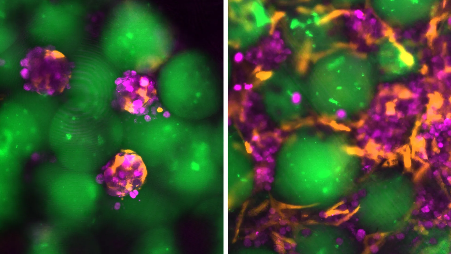
The researchers designed an injectable mixture containing liver cells and tiny hydrogel microspheres. These microspheres help keep the cells together and encourage connections with nearby blood vessels.
When densely packed, the spheres have properties that allow the material to behave like a liquid, making it possible to push the mixture through a syringe. Once inside the body, the material can recover its solid structure.
Hydrogel microspheres have previously been investigated for wound healing because they create spaces that allow cells to move through the material and generate new tissue. The MIT researchers adapted this approach so hepatocytes could establish a stable tissue graft following injection.
"What we did is use this technology to create an engineered niche for cell transplantation," Kumar says. "If the cells are injected in the absence of these spheres, they would not integrate efficiently with the host, but these microspheres provide the hepatocytes with a niche where they can stay localized and become connected to the host circulation much faster."
The injectable material also contains fibroblast cells -- supportive cells that improve hepatocyte survival and encourage blood vessels to grow into the newly formed tissue.
Working with Nicole Henning, an ultrasound research specialist at the Koch Institute, the team developed a technique for delivering the mixture with an ultrasound-guided syringe. Ultrasound can then be used after the procedure to track the implant and determine whether it remains stable over time.
Mini Livers Could Function in Different Parts of the Body
For the mouse experiments, the researchers placed the mini livers in fatty tissue within the abdomen. Future versions of the treatment could potentially be implanted in other locations, including the spleen or areas near the kidneys.
The liver cells do not necessarily need to be located next to the patient's liver. If the graft has enough room and a sufficient blood supply, the transplanted hepatocytes can perform functions similar to those of cells inside the liver itself.
"For a vast majority of liver disorders, the graft does not need to sit close to the liver," Kumar says.
Blood Vessels Help the Grafts Survive
In the mouse tests, the researchers injected the liver cells and microspheres into fatty tissue called the perigonadal adipose tissue. After being delivered, the cells gathered into a stable and compact structure.
Blood vessels gradually grew into the graft, creating a local supply of nutrients that helped keep the transplanted hepatocytes alive and functioning.
"The new blood vessels formed right next to the hepatocytes, which is why they were able to survive," Kumar says. "They were able to get the nutrients delivered right to them, they were able to function the way they're supposed to, and they produced the proteins that we expect them to."
The liver cells survived throughout the entire eight-week study and continued releasing specialized proteins into the animals' circulation. According to the researchers, that durability suggests the technique could potentially be developed into a longer-term treatment for liver disease.
A Potential Bridge to Liver Transplantation
The researchers envision several possible roles for the technology. For some patients, injectable liver grafts might provide an alternative to surgery. For others, they could temporarily supplement liver function while the patient waits for a donor organ.
"The way we see this technology is it can provide an alternative to surgery, but it can also serve as a bridge to transplantation where these grafts can provide support until a donor organ becomes available," Kumar says. "And if we think they might need another therapy or more grafts, the barriers to do that are much less with this injectable technology than undergoing another surgery."
The current approach would probably require patients to take immunosuppressive medications to prevent their immune systems from attacking the transplanted cells. The team is now investigating other possibilities, including the development of "stealthy" hepatocytes that could escape immune detection. Another option would be to use the hydrogel microspheres to release immunosuppressive drugs directly around the graft.
The research was funded by the Koch Institute Support (core) grant from the National Cancer Institute, the National Institutes of Health, the Wellcome Leap HOPE Program, a National Science Foundation Graduate Research Fellowship, and the Howard Hughes Medical Institute.
Story Source:
Materials provided by Massachusetts Institute of Technology. Note: Content may be edited for style and length.
Journal Reference:
- Vardhman Kumar, Joa Yun, Susanna K. Elledge, Nicole Henning, Katarzyna A. Grzelak, Ashley D. Westerfield, Amy Stoddard, Favour A. Oladimeji, Virginia Spanoudaki, Kasturi Chakraborty, Savan K. Patel, Heather E. Fleming, Christopher S. Chen, Sangeeta N. Bhatia. Image-guided injectable niche for hepatocyte transplantation. Cell Biomaterials, 2026; 2 (7): 100378 DOI: 10.1016/j.celbio.2026.100378
Cite This Page: