
Lab-grown mini brains may predict which Alzheimer’s treatments will work
- Date:
- July 22, 2026
- Source:
- Johns Hopkins Medicine
- Summary:
- Miniature brain models grown from patients’ cells revealed striking differences in how Alzheimer’s-related tissue responds to an antidepressant. The organoids and the particles they release could eventually guide more personalized treatments and provide new clues for diagnosing the disease.
- Share:
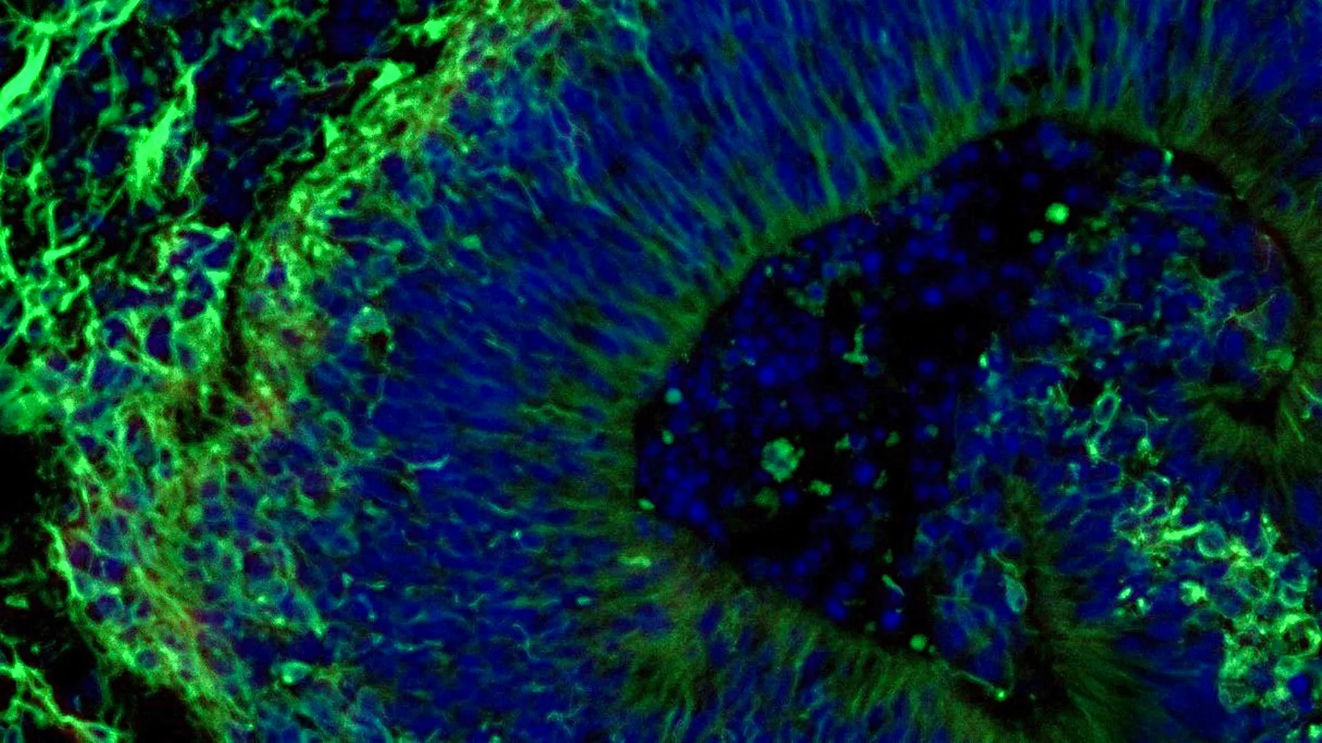
Scientists at Johns Hopkins Medicine have found new evidence that small clusters of brain tissue grown from the cells of people with Alzheimer's disease could help researchers predict how different patients may respond to medications used to manage psychiatric symptoms linked to the condition.
The research focused on laboratory-grown brain tissues called organoids. The findings add to growing evidence that these miniature brain models could eventually help scientists develop and select more precise treatments for specific groups of people with Alzheimer's disease. Alzheimer's is the most common form of dementia and affects more than 7 million Americans.
The team also discovered that the organoids release tiny particles called extracellular vesicles that carry cellular information. These particles may offer new biomarkers for diagnosing Alzheimer's disease and determining how far it has progressed.
The study, which received partial funding from the National Institutes of Health, was published in Alzheimer's & Dementia: The Journal of the Alzheimer's Association.
Mini Brain Models Could Support Personalized Care
"Our study suggests that large-scale, patient-derived brain organoids and the vesicles they secrete can help us stage Alzheimer's disease, investigate the mechanisms that drive it and assess how patient subgroups may respond to different treatments," says study leader Vasiliki Machairaki, Ph.D., associate professor of genetic medicine at the Johns Hopkins University School of Medicine.
There is currently no cure for Alzheimer's disease. However, selective serotonin reuptake inhibitors (SSRIs) are often prescribed to help manage neuropsychiatric symptoms such as anxiety, depression, and agitation. These symptoms affect nearly all patients, but responses to the medications vary widely, Machairaki says.
The Johns Hopkins researchers studied miniature models of the hindbrain, a region at the back of the skull that helps control essential functions such as breathing, sleep and heart rate. The team wanted to determine whether these models could reveal molecular signs showing whether the SSRI escitalopram oxalate might help reduce symptoms associated with Alzheimer's disease.
Turning Patient Blood Cells Into Brain Tissue
The researchers began with blood samples collected with permission from people with Alzheimer's disease at the NIH-funded Johns Hopkins Alzheimer's Disease Research Center.
They reprogrammed blood cells so that they returned to a stem cell-like state. These cells, called induced pluripotent stem cells, can develop into any type of cell in the body.
Using induced pluripotent stem cells from people with Alzheimer's disease and healthy individuals, the team created hindbrain organoids containing specialized brain cells, or neurons, that produce the neurotransmitter serotonin.
The cells were guided to organize themselves into small, pea-sized clusters of brain tissue that resemble the hindbrain. The study included hundreds of organoids representing individual patients with Alzheimer's disease as well as healthy participants. Machairaki believes it may be one of the largest brain organoid studies conducted so far in Alzheimer's research.
Alzheimer's Organoids Show Distinct Molecular Changes
The patient-derived organoids reproduced several important biological characteristics of Alzheimer's disease at the molecular level.
Compared with organoids created from healthy individuals, those grown from the cells of people with Alzheimer's showed differences in proteins involved in communication between brain cells, inflammation, and pathways associated with the disease.
The researchers then treated the organoids with escitalopram oxalate, a widely prescribed antidepressant.
In some organoids derived from patients, the medication increased proteins involved in serotonin signaling and communication between brain cells. These are pathways that antidepressants are designed to influence. Other organoids showed little or no molecular response.
"We used these organoids to model how some patients' tissue may respond to a commonly prescribed SSRI," Machairaki says. "On a large-scale level, our model may eventually be used to identify subgroups of patients, based on underlying molecular mechanisms, who are more likely to respond to certain drugs and thus help us to create precise, targeted treatments in the long run."
Tiny Vesicles May Reveal Drug Response
The team next investigated whether extracellular vesicles released by the organoids could serve as biomarkers of Alzheimer's disease or help researchers evaluate how tissue responds to treatment.
Before and after treating the organoids with escitalopram, the scientists examined proteins inside extracellular vesicles released by the patient-derived organoids and by healthy control organoids.
The vesicles contained proteins involved in essential brain activities, including communication between neurons, memory and the release of neurotransmitters.
Organoids grown from the cells of people with Alzheimer's showed clear changes in several disease-associated proteins. Levels of RAB3A, NSF and ATCAY were lower in the Alzheimer's organoids. These proteins play important roles in normal signaling between brain cells.
After escitalopram treatment, levels of some proteins increased in certain samples. The changes were especially noticeable in proteins connected to serotonin signaling and synaptic pathways that antidepressants target.
Some organoids displayed strong molecular responses, while others showed little or no change. According to Machairaki, this variation raises the possibility that extracellular vesicles from brain organoids could eventually help identify which patients are most likely to benefit from a particular treatment.
Building More Realistic Brain Organoids
Machairaki plans to develop more advanced organoids that contain immune cells and vascular-like networks that imitate blood vessels. Adding these features could make the tissues more similar to living human brain tissue.
With additional research, she hopes extracellular vesicles might one day function as a type of liquid biopsy. Such a test could potentially help diagnose Alzheimer's disease, determine its stage, and identify a patient's particular disease subtype.
Machairaki emphasized that the current study represents an early step toward that goal.
In addition to Machairaki, scientists who contributed to this work include Rachel Boyd, Daiyun Dong, Ram Sagar, Waqar Ahmed, Xenia Androni, Paul Rosenberg, Constantine Lyketsos and Kenneth Witwer from Johns Hopkins, Anton Iliuk from Tymora Analytical Operations and Anton Porsteinsson from University of Rochester School of Medicine and Dentistry.
Funding for this study was provided by the National Institutes of Health (T32 AG058527, R01AG052510, P30AG066507, 1RF1AG083801, AGR01054771, AGR01050515, AGR01046543 and AGR01071522), the Paul G. Allen Frontiers Foundation and the Richman Family Precision Medicine Center of Excellence in Alzheimer's Disease at The Johns Hopkins University.
No authors declare a related conflict of interest under Johns Hopkins University policies.
Story Source:
Materials provided by Johns Hopkins Medicine. Note: Content may be edited for style and length.
Journal Reference:
- Rachel J. Boyd, Daiyun Dong, Ram Sagar, Anton Iliuk, Waqar Ahmed, Xenia Androni, Anton P. Porsteinsson, Paul B. Rosenberg, Constantine G. Lyketsos, Kenneth W. Witwer, Vasiliki Mahairaki. Proteomic profiling of brain organoids and extracellular vesicles identifies early Alzheimer\'s disease biomarkers and drug response heterogeneity. Alzheimer\'s, 2026; 22 (4) DOI: 10.1002/alz.71273
Cite This Page: