New brain imaging breakthrough reveals clues to Parkinson’s
A rapid “zap-and-freeze” method exposes hidden steps in brain communication that may help explain nonheritable Parkinson’s.
- Date:
- December 1, 2025
- Source:
- Johns Hopkins Medicine
- Summary:
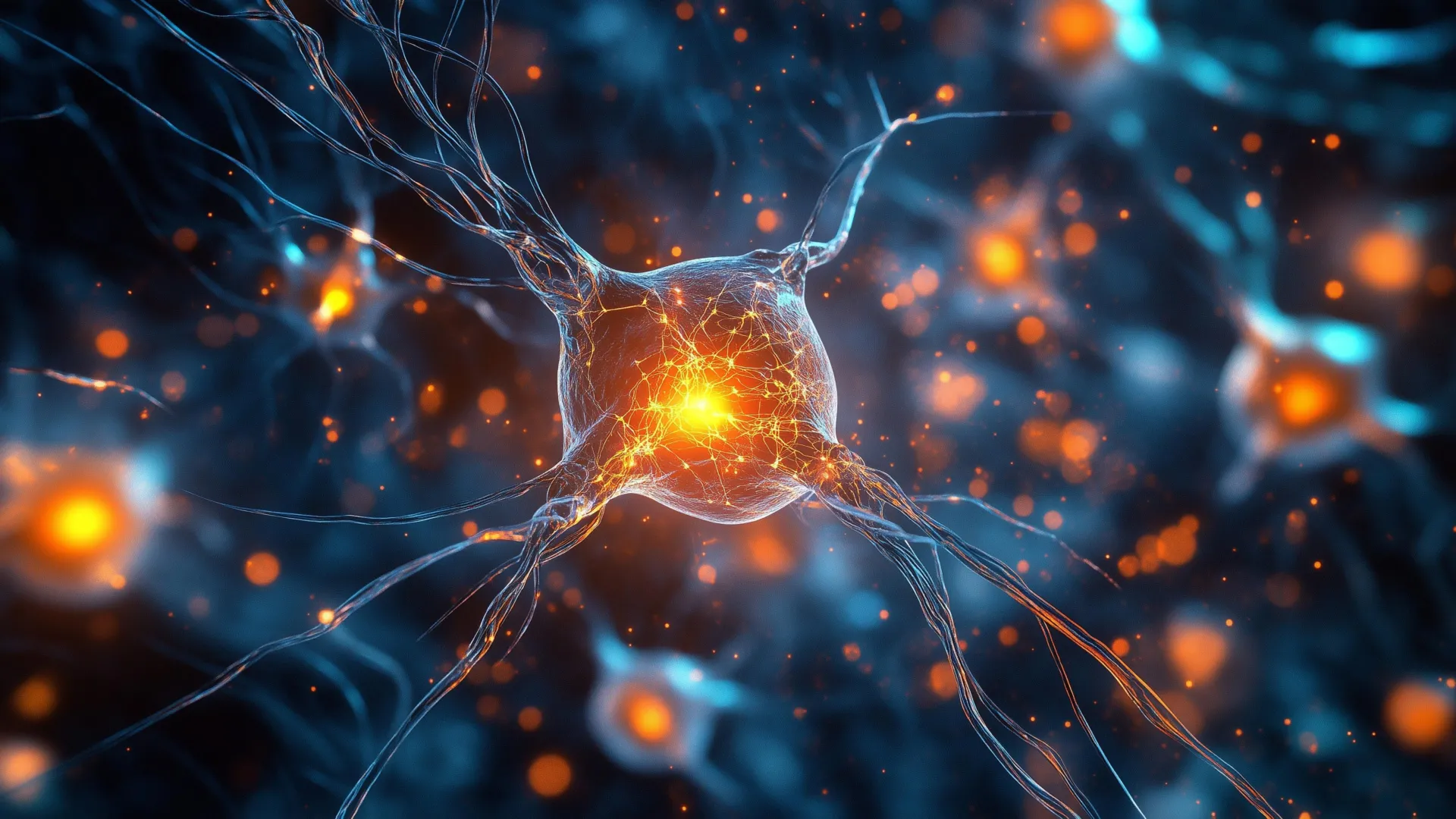
- A high-speed “zap-and-freeze” method is giving scientists their clearest view yet of how brain cells send messages. By freezing tissue at the instant a signal fires, researchers revealed how synaptic vesicles behave in both mouse and human neurons. These insights could help explain why most Parkinson’s cases emerge without inherited genetic changes. The technique may also point to promising new research paths for therapy development.
- Share:
Researchers at Johns Hopkins Medicine report that they have successfully used a "zap-and-freeze" method to capture rapid communication between brain cells in living tissue from both mice and humans. The approach allowed them to observe interactions that normally happen too quickly to track.
According to the team, the findings, published Nov. 24 in Neuron and supported by the National Institutes of Health, may help uncover the underlying biological causes of nonheritable forms of Parkinson's disease.
Sporadic Parkinson's cases represent the majority of diagnoses, the Parkinson's Foundation notes. These cases involve disruptions in the synapse, the tiny site where one neuron passes a signal to another. Because this junction is so small and its activity unfolds rapidly, it has long been challenging to study in detail, says Shigeki Watanabe, Ph.D., an associate professor of cell biology at Johns Hopkins Medicine and the senior author of the study.
"We hope this new technique of visualizing synaptic membrane dynamics in live brain tissue samples can help us understand similarities and differences in nonheritable and heritable forms of the condition," Watanabe says. He adds that the technique could eventually guide the development of therapies for this neurodegenerative disorder.
How Healthy Synapses Move Messages
In a healthy brain, synaptic vesicles act as tiny packages that carry chemical messages from one neuron to the next. This exchange is essential for learning, memory formation and the processing of information. Understanding how vesicles behave under normal conditions is key to identifying where communication begins to fail in neurological diseases, Watanabe says.
Watanabe previously helped design the zap-and-freeze approach to visualize fast changes in synaptic membranes (these results were published in 2020 in Nature Neuroscience). The method uses a brief electrical stimulus to activate brain tissue, followed immediately by rapid freezing. This preserves the exact positions of cellular structures for later viewing with electron microscopy.
In earlier work published in Nature Neuroscience this year, Watanabe applied the method to genetically engineered mice to investigate the role of a protein called intersectin. The study demonstrated how intersectin helps maintain synaptic vesicles in a specific location until they are ready to be released and activate a neighboring neuron.
Testing the Technique in Human Brain Tissue
For the latest study, the team examined samples from normal mice and compared them with living cortical brain tissue obtained, with permission, from six people undergoing epilepsy surgery at The Johns Hopkins Hospital. These surgeries were necessary to remove hippocampal lesions.
Collaborating with Jens Eilers and Kristina Lippmann of Leipzig University in Germany, the researchers first confirmed that zap-and-freeze worked reliably in mouse tissue by observing calcium signaling, which is the trigger that prompts neurons to release neurotransmitters.
They then used the technique to stimulate mouse neurons and captured the moment when synaptic vesicles fused with the cell membrane and released their chemical messengers. The researchers also documented how the cells retrieved and recycled vesicles afterward, a process known as endocytosis.
When the team applied zap-and-freeze to the human tissue samples, they found the same vesicle recycling steps occurring in human neurons.
Key Protein Found in Both Mouse and Human Brains
In both species, the researchers identified the presence of Dynamin1xA, a protein required for ultrafast synaptic membrane recycling, at the locations where endocytosis is believed to take place. This similarity suggests that the mechanisms observed in mice accurately reflect those in humans.
"Our findings indicate that the molecular mechanism of ultrafast endocytosis is conserved between mice and human brain tissues," Watanabe says. He notes that this strengthens the value of using mouse models to study human brain biology.
Looking ahead, Watanabe hopes to apply the zap-and-freeze method to brain tissue collected, with permission, from individuals with Parkinson's disease who are undergoing deep brain stimulation procedures. The goal is to observe how vesicle dynamics may differ in affected neurons.
Funding for the study was provided by the National Institutes of Health (U19 AG072643, 1DP2 NS111133-01, 1R01 NS105810-01A1, R35 NS132153, S10RR026445), Howard Hughes Medical Institute, Kazato Foundation, American Lebanese Syrian Associated Charities, Marine Biological Laboratory, Leipzig University, Roland Ernst Stiftung, Johns Hopkins Medicine, Chan Zuckerberg Initiative, Brain Research Foundation, Helis Foundation, Robert J Kleberg Jr and Helen C Kleberg Foundation, McKnight Foundation, Esther A. & Joseph Klingenstein Fund, and the Vallee Foundation.
Contributors to the research included Chelsy Eddings, Minghua Fan, Yuuta Imoto, Kie Itoh, Xiomara McDonald, William Anderson, Paul Worley and David Nauen from Johns Hopkins, along with Jens Eilers and Kristina Lippmann from Leipzig University.
Story Source:
Materials provided by Johns Hopkins Medicine. Note: Content may be edited for style and length.
Journal Reference:
- Chelsy R. Eddings, Minghua Fan, Yuuta Imoto, Kie Itoh, Xiomara McDonald, Jens Eilers, William S. Anderson, Paul F. Worley, Kristina Lippmann, David W. Nauen, Shigeki Watanabe. Ultrastructural membrane dynamics of mouse and human cortical synapses. Neuron, 2025; DOI: 10.1016/j.neuron.2025.10.030
Cite This Page: