Scientists capture most detailed look inside DNA droplets
- Date:
- December 8, 2025
- Source:
- Howard Hughes Medical Institute
- Summary:
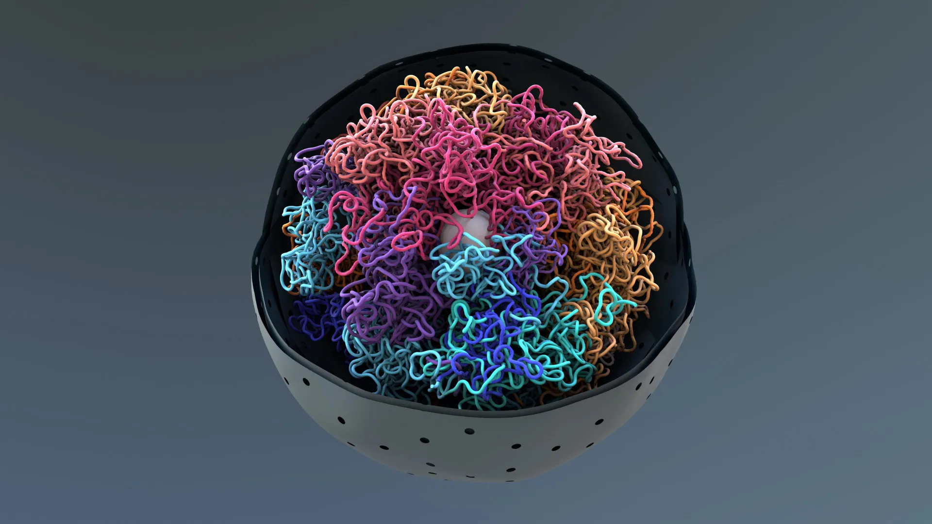
- High-resolution imaging has revealed the internal layout of chromatin condensates, showing how DNA fibers fold and interact within these droplet-like structures. The findings connect molecular architecture to the broader behaviors of these droplets in cells.
- Share:
Inside every human cell, an extraordinary feat of molecular organization takes place. Roughly six feet of DNA has to be packed into a nucleus that is only about one-tenth the width of a human hair, yet the DNA must remain accessible enough to carry out essential functions.
To make this possible, DNA coils around proteins to form nucleosomes. These nucleosomes connect like beads on a string and fold into chromatin fibers. The fibers are then compacted even more tightly to fit inside the nucleus.
Discovering How DNA Reaches Its Most Compact Form
For many years, researchers did not know how this extra level of chromatin compaction occurred. In 2019, HHMI Investigator Michael Rosen and his team at UT Southwestern Medical Center reported that lab-made nucleosomes naturally cluster together into membrane-less droplets called condensates. They found that this process occurs through phase separation, a phenomenon similar to oil droplets forming in water, and they believe it reflects how chromatin condenses inside living cells.
Chromatin condensates are made of hundreds of thousands of fast-moving molecules. When they come together, they display emergent properties that do not exist in the molecules individually. These group behaviors determine how condensates form and how they maintain their physical characteristics.
To understand these properties in detail, scientists needed to observe chromatin fibers and nucleosomes deep inside the droplets.
Rosen's group, working with HHMI Investigator Elizabeth Villa at the University of California, San Diego; Rosana Collepardo-Guevara at the University of Cambridge; and Zhiheng Yu at HHMI's Janelia Research Campus, has now achieved that goal.
High-Resolution Imaging Reveals Droplet Structure
Using advanced imaging tools at Janelia, the researchers captured the most detailed views to date of how molecules are arranged inside synthetic chromatin condensates. These images provide a direct look at how chromatin fibers and nucleosomes are packaged within the droplet-like structures. The same imaging methods were also applied to examine chromatin inside actual cells.
By combining these images with computer simulations and light microscopy, the team analyzed the molecular structures and interactions within the synthetic condensates. This allowed them to begin uncovering how droplets form and how they behave.
One important discovery was that the length of linker DNA between nucleosomes influences the overall arrangement of the structures. That arrangement determines how chromatin fibers interact and shapes the network inside the condensates.
These features clarified why some chromatin fibers undergo phase separation more easily than others and why condensates built from different chromatin types have distinct material properties. The researchers also found that synthetic condensates closely resemble compacted chromatin found in cells.
"The work has allowed us to tie the structures of individual molecules to macroscopic properties of their condensates, really for the first time," Rosen says. "I'm certain that we're only at the tip of the iceberg -- that we and others will come up with even better ways of developing those structure-function relationships at the meso (intermediate) scale."
A Broader Framework for Understanding Condensation
The findings extend well beyond chromatin. The approach offers a model for studying many kinds of biomolecular condensates, which are membrane-less droplets involved in essential cellular tasks from gene regulation to stress responses.
Understanding how these structures assemble and operate may also shed light on what happens when condensation is disrupted, a problem thought to contribute to various diseases from neurodegenerative disorders to cancer.
"By doing this research, we will better understand how abnormal condensation could lead to different diseases and, potentially, that could help us develop a new generation of therapeutics," says Huabin Zhou, a postdoctoral scientist in the Rosen Lab and the lead author of the new research.
Story Source:
Materials provided by Howard Hughes Medical Institute. Note: Content may be edited for style and length.
Journal Reference:
- Huabin Zhou, Jan Huertas, M. Julia Maristany, Kieran Russell, June Ho Hwang, Run-Wen Yao, Nirnay Samanta, Joshua Hutchings, Ramya Billur, Momoko Shiozaki, Xiaowei Zhao, Lynda K. Doolittle, Bryan A. Gibson, Andrea Soranno, Margot Riggi, Jorge R. Espinosa, Zhiheng Yu, Elizabeth Villa, Rosana Collepardo-Guevara, Michael K. Rosen. Multiscale structure of chromatin condensates explains phase separation and material properties. Science, 2025; 390 (6777) DOI: 10.1126/science.adv6588
Cite This Page: